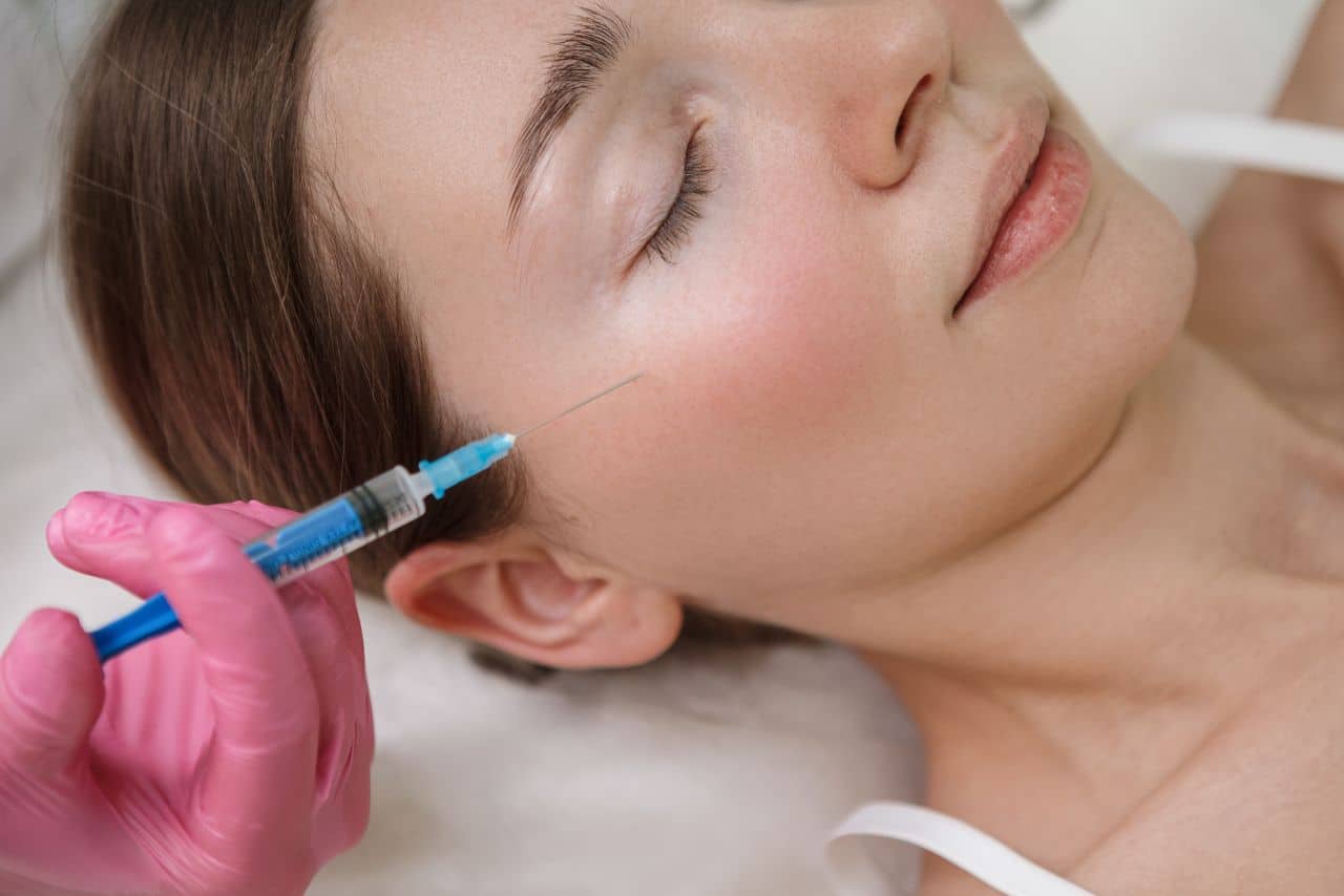
Complications from dermal fillers are the bane of aesthetic medicine. It is known to a certain degree by most medical professionals that the complications are mostly mild and transient. It is also known that the more severe complications are the most dreaded because of their more permanent nature. Therefore, any successful aesthetic practitioner must be aware of these side effects and their intrinsic nature. In light of the potential severity of some of these side effects, this article aims to address many of these complications in respect to their time of occurrence and the methods to tackle them.
Considerations when using dermal fillers
There are a multitude of simple and practical ways that the aesthetic practitioner can take to protect their patients. First, a well-regulated and stringent aseptic technique should be taken into consideration when performing procedures as minimally invasive as dermal filler injections. It is essential to minimize the risks of infection. Steps from hand washing, using sterile packs, swabs, and gloves should absolutely be followed. Ensure that the skin is properly cleansed with chlorhexidine, not alcohol, as there is some evidence suggesting that the former provides a longer anti-microbial action.
For deeper tissues that require volume augmentation, they may require multiple needles or cannulas for injection. Practitioners should not touch or wipe their needle with a swab prior to injection. It is neither desirable nor sensible to contaminate a sterile object. Indeed, it is against standard protocol; if a sterile surface has been in contact with a non-sterile surface, the once sterile surface is now considered contaminated and should not be used. If you have touched needle or cannula or let them touch anything other than the area of treatment, you must change it.
Injection techniques are also an important part of preventing complications specific to the use of fillers, such as bruising and swelling. Slow and small aliquot deposition techniques are preferable in this respect It is also more comfortable for the patient, as they will feel less pain.
Addressing Complications
Minimizing risks
It is necessary for the injector to have comprehensive knowledge on the facial vascular anatomy, including arteries, and high-risk areas. The exact location of parts of the anatomy can also vary from patient to patient, so keep that in mind. In terms of anatomy, the angular artery runs just lateral to the nasolabial fold. On the other hand, the angular vein is near the eye. It is formed from both the frontal and supraorbital vein, and it runs obliquely inferior to the side of the root of the nose and to the level of the lower margin of the eye socket, where it forms the anterior facial vein. The branches of the facial vein are typically found along the marionette fold, and this exact area is a very common site for bruising incidences. With that said, a good alternative to the needle is using the cannula in this area. When treating the lip area, remember that the labial artery courses deep into the body of the lip, and any injection along the wet/dry border is likely to also have bruising.
Early complications
Early complications are generally classified when they occur between zero to 14 days after the procedure. Most of them are related to the technique of injection instead of the filler content. Both bruising and swelling are extremely common after injection, and because of this, the patients should be notified to consider rescheduling any social events that are within the next few days. It is difficult to ascertain the exact duration of these side effects due to the fact that they vary from case to case. However, to reduce their incidence, use the needle with the highest gauge to minimize trauma, and consider using blunt tipped micro cannulas for high-risk areas.
In terms of swelling, technique plays a major role in its occurrence, but you must also consider the hygroscopic properties of the dermal filler material. When treating the lips, it is important to note that any swelling will be considerably more apparent. Inform your patients about this reality and ask for their opinion to know if they find it acceptable. Usually no more than 1ml of dermal filler material is used in lip enhancement during the initial stages of treatment. To reduce the impact of swelling, treatment should occur in two-week intervals. Also, consider the use of ice packs and oral steroids, as they are well known to mitigate the pathophysiology behind swelling in this case. Again, patients may want to reschedule social events for at least one if they are uncomfortable with the idea of their possibly swollen lips being visible. For severe bruising, many treatment options are available if the patient desires a faster resolution. One such option is a vascular laser (i.e. pulsed dye laser) treatment. Lumenis One IPL is one such treatment and has been demonstrated to be an effective treatment.
The most serious complication for dermal filler injections is inadvertent intravascular injection because it can lead to tissue necrosis and potentially irreversible scarring. This complication may be glossed over by some practitioners due to its relatively rare occurrence, but it is absolutely mandatory to know the course of immediate management if such a case is ever present. In addition to having excellent knowledge about the surface anatomy prior to the injection, it is also important to know that there are areas that are considered dangerous. For example, the glabella is a watershed area due to the supratrochlear and supra orbital arteries. These arteries are end arteries from the internal carotid artery, which means that there will be minimal collateral circulation in the event of an intravascular injection that occludes the blood flow to the area. Moreover, the lack of distance between the skin and periosteum of the frontal bone should add to the concern. Deep frown lines should be treated with more precaution, as patients with them are the type that are the most at risk. They can also be harder to satisfy, as treating deep frown lines often yield minimal improvements.
The steps required to handle these patients start with pre-treatment. Effectively communicate to them that their static lines will progressively improve. Emphasize on the duration of treatment sessions and formulate an easy-to-comprehend treatment plan. Make sure that patients understand that their imperfection will not be completely eliminated but only improved to a substantial degree. Have proper documentation via photography of pre- and post-treatment. The glabellar arteries can be so fragile that they can be compressed if excessive filler material is deposited. This can also lead to signs of tissue ischemia and eventually skin necrosis, which will be elaborated later on.

The nasolabial fold can be regarded as an area suitable for less-experienced cosmetic practitioners. The facial artery, which is a branch of the external carotid artery, lies just lateral to this fold. It continues on to become both the upper and lower labial branches, which are lateral to the oral commissure. At the base of the fold, there is commonly a branch that crosses medially at the nasal ala. Any intravascular injection into the artery has a high chance of causing an embolism that occludes the blood supply to the nose. Compression can also easily occur at this area. Any post-procedural pain is not acceptable, nor should it be taken lightly. Provide patients with an emergency contact and be sure to notify patients of early symptoms of ischemia.
Rhinoplasty has continued to remain a popular procedure since its inception. However, they are one of the most technically demanding procedures and require expert judgement from specialists. There is a high risk of skin necrosis. Nasal tip necrosis as a result of occluding post filler is most likely associated with compression of the vessels in this area and not direct intravascular injection. Embolization has also been known to occur proximally at the area of the nasal bone, where it can cause permanent blindness as it travels to the retinal artery. 9 With these in mind, it is vital for you, as the injector, to inject slowly and gently while constantly moving the tip of the needle. Do not attempt bolus injections. The literature may advocate aspiration of the needle prior to injection, but practical reasons, such as the needle tip being in different positions during aspiration and after aspiration, may compromise the usefulness of this practice. Always watch for signs of ischemia, with pain and pallor particularly important. If these early signs manifest, treat patients immediately with a gentle massage, heat, topical glyceryl trinitrate, and most importantly, hyaluronidase. For cases of embolization, hyaluronidase should still be used. Make an emergency referral to a plastic surgeon.
Blindness from dermal filler injection is highly preventable, but its occurrence in recent years has caused some concern among users. These cases, however, are primarily from fat injections but, theoretically, it can happen in any other dermal filler material. The majority of the arterial blood supply to the face is derived from the external carotid artery. The exceptions are the glabella, supratrochlear, and the supraorbital arteries. These come from the internal carotid artery instead, and because of this, they communicate with the retinal artery. There are also multiple sites of anastomoses between the branches of the external and internal carotid artery. They are the base of the nose, the temple, and the tear trough. These are highly specific and dangerous areas that you must take note of prior to injection.
Be vigilant to note early signs of infections, such as erythema and tenderness, as they can occur with any foreign material deposited into the skin. If they are present, prescribe macrolide antibiotics, such as clarithromycin at 500mg BD or ciprofloxacin at 750mg BD, for at least two weeks.
Medium term complications
Medium term complications can sometimes be relatively harder to treat. Palpable nodules are frequent if there are incidences of overcorrection. They can be massaged to fall in line with the contours of the surrounding tissue if they are small and non-inflamed. Signs of infection should prompt early and judicious use of antibiotics. Use hyaluronidase if there is a need for reversal of the treated area.
The common treatment areas, such as the lip, tear trough, and cheeks, can frequently have contour irregularities post-treatment. In the lip region, the orbicularis oris can further exacerbate this issue through its muscular contraction. Furthermore, hyaluronic acid dermal fillers can, if the depth of injection is too superficial, cause a bluish discoloration of the skin. This is known as the Tyndall effect and is due to the refraction of light off of the hyaluronic acid particles. This discoloration can last for a few years. The pattern seems to be that the more superficial the dermal filler is placed, the longer it will likely last. Hyaluronidase is the appropriate solution for these cases. However, hyaluronidase is not explicitly indicated for dissolving hyaluronic acid dermal fillers. Also, it is manufactured using animal parts; therefore, there is a risk of a hypersensitivity reaction. Patch testing on the forearm may be a safe choice prior to hyaluronidase administration. Dissolving 0.1ml of hyaluronic acid dermal fillers usually require about 10 to 15 units of hyaluronidase, although this depends on the exact filler used. Start with small aliquots before full injection. Have the hyaluronidase solution prepared on hand prior to dermal filler injection.
Late complications
In contrast with permanent dermal fillers, the late complications of temporary dermal fillers are few and far between. Foreign body granulomas occur at a rate of 0.01% to 1.0% of all dermal filler injections. They typically occur between six to 24 months. It is now universally accepted that biofilms play a central figure in producing this late complication. Biofilms are basically low pathogenic bacteria that has accumulated onto the implanted dermal filler material. The foreign body functions as a colonization target, and the bacteria subsequently secretes a protective layer of polysaccharides that makes it impermeable to the immune system and antibiotics. In two-thirds of the cases, the culprit bacteria seem to be Staphylococcus aureus, Staphylococcus epidermidis, Pseudomonas aeruginosa, and the Enterococcus species. They are also crucial in producing many subacute and chronic infections. Biofilms more commonly result from permanent dermal fillers, such as silicone or polyacrylamide materials, than more temporary agents. Biofilms can also result in other clinical responses, including abscesses, cellulitis, sepsis, or nodules. A common treatment tactic is to administer a high dose of antibiotics for late nodules to impair biofilm resistance. Most of the time, however, surgical removal (e.g. excision) is required for complete resolution. Hyaluronic acid offers a distinct advantage in this regard, as hyaluronidase can be used in place of surgery for reversal. Common antibiotic regimes are taking ciprofloxacin 750mg BD or clarithromycin 500mg BD for four weeks. Advise patients against undergoing vigorous exercise, as it makes them more prone to tendinopathy.
Conclusion
As with many other surgical or medical procedures, complications may seem inevitable. Nonetheless, soft tissue augmentation with dermal fillers continue being among the most commonly performed cosmetic procedures. If they are performed with by experienced practitioners who show due diligence, it can be a highly effective and extremely well-tolerated procedure. As the demands from patients increase, the types of fillers continue to advance and expand. It is imperative, then, that practitioners have up-to-date knowledge on these products so that they can handle the potential complications. Although the overall rates for complications from dermal fillers are low, they can still occur, and when they do, it demands immediate recognition and management because the consequences can be dire. To best minimize these adverse events, you must have elaborate knowledge of the facial anatomy, good judgement to choose the product to be used, and the ability to perform the proper injection technique(s).
