
The earlobes are important facial features that form an integral part of the overall facial aesthetic. Because of this, deformed earlobes (such as asymmetrical earlobes or earlobes that are too large) are a major cause of concern. This article will discuss the anatomy of the earlobes, the common conditions associated with the earlobes, and the non-surgical procedures used to treat them.
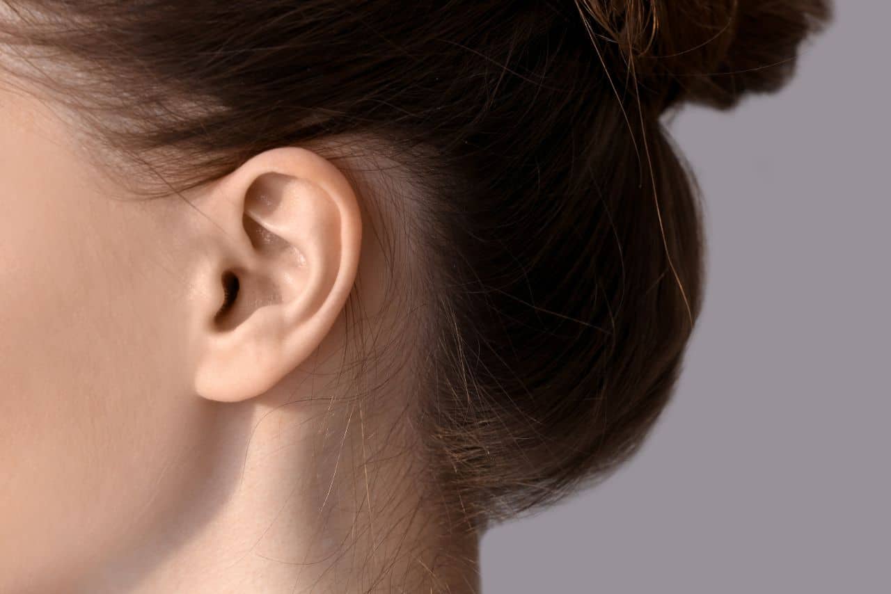
Before treating the earlobes, it is important to understand the external ear anatomy. The external ear consists of the ear canal and pinna (also referred to as the auricle). The auricle is the visible part of ear that is comprised of skin-covered cartilage. The components of the auricle include the concha, tragus, lobule, antihelix, and helix. The human earlobe is made of adipose connective tissues and tough areolar, meaning that it lacks the elasticity and firmness of the auricle.
Essentially, there are 2 forms of earlobes: unattached (where the lower lobe is not connected to the cheek) and attached (where the lower lobe is connected to the cheek). With its extensive blood supply and innervation system, understating the anatomy of this area is essential to minimizing the risk of complications during reparation procedures.
Sensory innervation of the external ear
In order to understand how to anesthetize the ear during remodeling procedures, it is essential to understand the anatomy of its nerves. There are 4 sensory nerves that supply the ear, including the auriculotemporal nerve, the auricular branch of the vagus nerve, the lesser occipital nerve, and the great auricular nerve. The auriculotemporal nerve is known to innervate the anterior superior third of the ear, while the auricular branch of the vagus nerve innervates the concha and external auditory canal floor. Additionally, the lesser occipital nerve innervates the posterior surface of the superior third of the external ear, whereas the great auricular nerve innervates the lower two thirds of the posterior and anterior external ear.
Blood supply to the ear
There are 3 branches in the external carotid artery that provide blood to the ear. This includes the anterior auricular branch of the superficial temporal artery, the posterior auricular artery, and the occipital artery. Note that veins run along with these arteries. To ensure continuous supply of blood to the ear during medical procedures, practitioners should have a good knowledge of the ear’s blood supply.
Earlobe Concerns
Earrings and ear piercings may cause ear trauma, potentially resulting in keloid scarring, enlarged ear piercing holes, and split earlobes.
1. Enlarged piercing hole
Over the past few years, there has been an increase in the number of individuals who wear enlarged plugs in their earlobes. This process stretches the ears, and is known as gauging. It is a piercing hole-enlarging method in which expandable gauge earrings are used to stretch the hole. Using a tapering device, the size of the gauge can be increased from 1mm to over 2cm. To reduce the size of this hole, wearers may gradually reverse the size of their gauging earrings. However, overly-stretched piercing holes are not fully reversible.
2. Split earlobe
Split earlobes are usually caused by heavy, large earrings, as these may put extra weight on the earlobe. Furthermore, catching earrings while combing the hair could lead to ear trauma. There are 2 major types of traumatic split earlobe: incomplete and complete. Complete traumatic split earlobe is characterized by a fully torn piercing hole, whereas incomplete traumatic split earlobe involves a slightly torn piercing hole.
Treatment Options
Treatments should be individualized for every patient. Carefully evaluate each case to determine the most appropriate treatment. Keep in mind that when it comes to treating earlobes, 1 size does not fit all.
1. Repairing enlarged piercing holes
It is recommended to perform an L-plasty for big ear piercing holes, and a Y-plasty for piercing holes that are very big. The treatment is chosen based on the amount of tissues available in the earlobe. Elliptical excision is commonly used to repair a very small piercing hole.
2. Earlobe reduction
To prevent scarring or notches on the free border, excise the earlobe to remove excess tissues. Prior to the procedure, the doctor should ask the patients about their preferred size of earlobe. Mark the region using a white marker. Then draw a line connecting the tragus with the white mark. Draw another line on the earlobe connecting the tragus with the lateral end of the white mark. After measuring the lengths of these 2 lines, draw a triangle on the second line. Make sure that the base of the triangle is the same size as the length of both lines. The triangle cut will reduce the length of the 2nd line to the same as the 1st line. Perform the excision as marked. Stitch the triangle to make the 2nd line shorter. Using vertical mattress and deep dermal stitches, join the 1st and 2nd lines.
3. Incomplete split earlobe repair
It is recommended to repair incomplete split earlobes using an L-plasty. Due to the risk of earlobe elongation, do not cut and stitch the skin near the edge of the split. Some patients may find an elongated earlobe unacceptable.
4. Split earlobe repair
Split earlobes can be repaired using several techniques. Some of them may involve the reconstruction of the piercing hole. Patients are advised to wait for 6 months before re-piercing their ears. To avoid the split from occurring again, piercing should be performed at around 2mm lateral or medial to the scar.
Generally, there are 2 methods of repairing a split earlobe, which are broken-line and straight-line repair. Broken-line repair (also referred to as L-plasty or the lab-joint technique) forms a Z-shaped wound to prevent the scar from shrinking. At the same time, it prevents notch effects on the free border of the earlobes. While straight-line repairs are simple, they may not deliver aesthetically acceptable results. Simple line repair often causes the contraction of the scar. Moreover, a notch may form at the free border of the earlobe.
Earlobe repair procedure
Earlobe repairs should be performed using a local anesthesia. The details of the procedure are as follows.
- When performing Y-plasty and L-plasty, excise the areas as marked using a pair of Iris scissors or scalpel. Start from the free border all the way to the apex of the earlobe cleft. Make sure that the back of the earlobe is cut to the same shape as the front. Then, insert the flap. Start from the anterior aspect. Any discrepancy will be transferred to the posterior aspect. This will eliminate any irregularity in the free border.
- When performing L-plasty, the excision lines should be drawn at the highest point on the cleft (either side is fine) before the edges gently curve in. Otherwise, there will be a groove along the repair line. This will create a split earlobe with a notched border.
- Using vertical mattress technique, stitch to the desired shape with the non-absorbable sutures at the back and front of the earlobe. Always use the vertical mattress technique to avoid the inward rolling of the scar. Otherwise, the scar may appear more prominent.
- Administer around 2ml of local anesthetic (2% lidocaine or 2% xylocaine with adrenaline) to the junction of the cheek and earlobe superficially via subdermal injection. This helps to reduce the risk of injuring the facial nerve. The anesthetic should be injected from the lowest end of the junction all the way up to the intertragic notch at both the front and back of the junction. As an alternative, use the anesthetic solution (without adrenaline) to infiltrate the earlobe until it appears pale and firm. This will make it easier to excise.
Warnings and precautions
When choosing the right earlobe repair procedure, consider the amount of tissues available and the preference of each patient. In order to prevent further complications, the following precautions should be taken.
- These procedures are associated with a risk of hematoma, bruising, and bleeding. Before undergoing this treatment, patients should not take any blood-thinning agents such as ibuprofen, aspirin, vitamin C and E, and alcohol. Make sure that the stiches are properly tightened.
- If there is an infection, patients should avoid touching the treated area with water. They are advised not to wash their hair 2 to 3 days after the procedure. Instead, patients should wash their hair the morning before undergoing the procedure.
- Patients with skin types V to VI are more susceptible to scarring. If patients have had hypertrophic scars or keloids, they are at a higher risk of developing keloid scarring. Patients should be informed of the risk of scarring prior to the treatment.
- Measure the marks on both earlobes to ensure that they have the same shape, length and angle. Sit the patient up to check. Before the procedure, patients should be warned of the risk of getting earlobes that do not appear exactly the same. This is due to the difference in the amount of tissues available in each earlobe. When performing earlobe reduction, the doctor should measure the marks on both earlobes to ensure consistent results.
- Avoid applying any pressure or stress to the treated area, as this could loosen the stitches. Patients are advised to sleep on their back after the procedure. In addition, they should avoid any activities that can potentially pull the ear (e.g. contact sports).
- Be sure to tighten all stitches. Failure to do so may result in bleeding after the procedure. Plus, the wound will not close properly if the stiches are not tightened.
- Triamcinolone (TCN) may result in an anaphylactic reaction (specifically, type 1 anaphylactic reaction). Anaphylactic reactions are usually triggered by Immunoglobulin E (IgE) antibodies. There are several allergens in triamcinolone, including succinate, carboxymethyl cellulose (CMC) and steroid. The medical history of patients should be assessed for the presence of allergies (in particular, allergies to steroids).
- Before stitching, check the back of the earlobe to ensure that the shape is correct.
Conclusion
With so many options available, choosing the right treatment can be tricky. There are a lot of factors that needs to be considered when determining the most appropriate technique. As a general rule, Y-plasty technique should be used for large hole repair and earlobe reduction, while L-plasty technique is usually indicated for split earlobe repair.

About the Author: Doris Dickson is a specialist writer for Health Supplies Plus, focusing on the aesthetic medicine industry. She diligently researches cosmetic treatments and products to provide clear, concise information relevant to licensed medical professionals. Her work supports Health Supplies Plus’s commitment to being a reliable informational resource and trusted supplier for the aesthetic community.
Disclaimer: The content provided in this article is intended for informational purposes only and is directed towards licensed medical professionals. It is not intended to be a substitute for professional medical advice, diagnosis, or treatment, nor does it constitute an endorsement of any specific product or technique. Practitioners must rely on their own professional judgment, clinical experience, and knowledge of patient needs, and should always consult the full product prescribing information and relevant clinical guidelines before use. Health Supplies Plus does not provide medical advice.