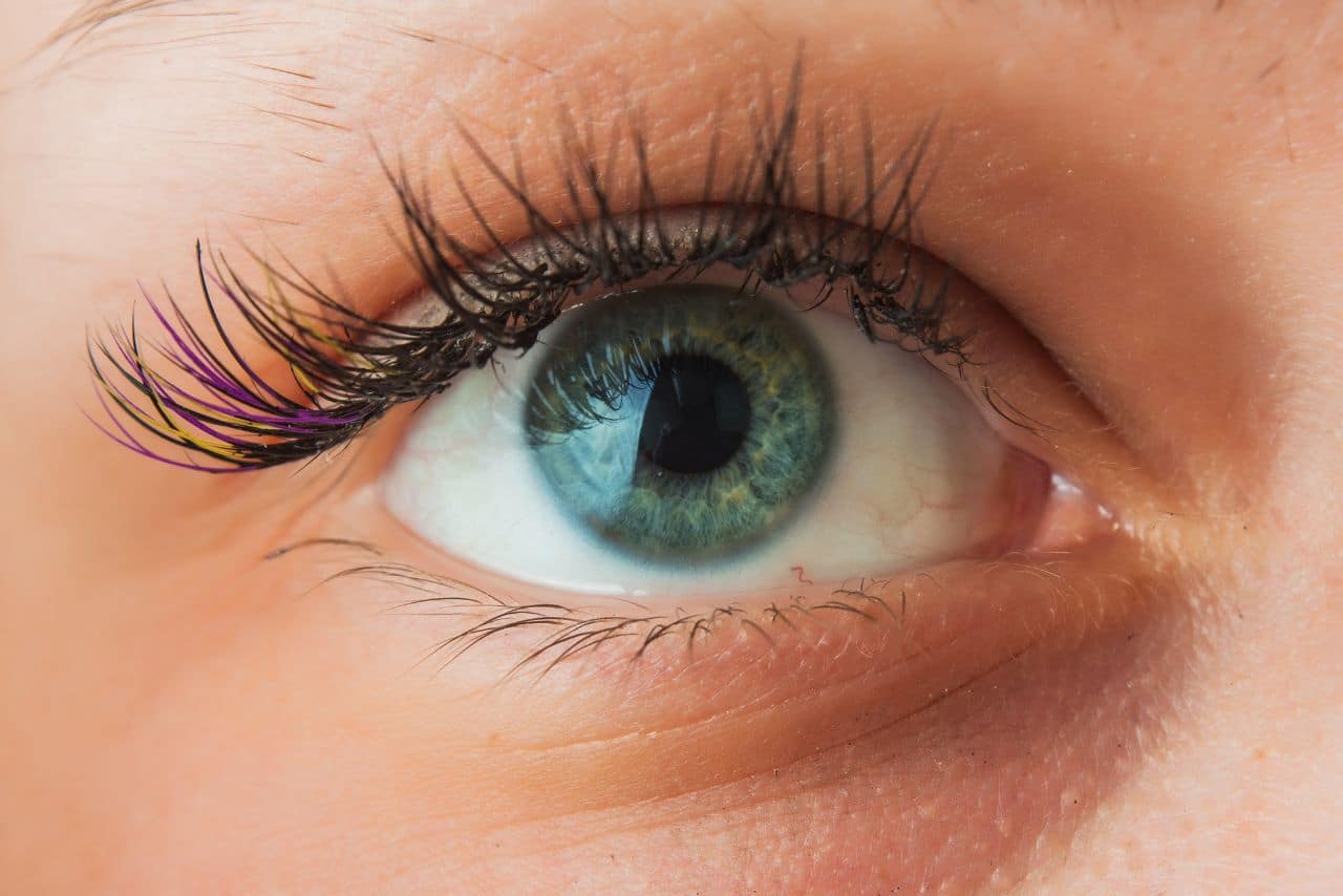
Eyelid dermatochalasis
Periocular dermatochalasis is characterized by eye bags and an excessively loose skin. It often affects the upper eyelids, resulting in the loss of elasticity in the skin fold. Apart from that, it also causes hooding (i.e. where skin fold drops down and outwards).
Redundant or excessive subcutaneous tissue and upper eyelid skin may be formed due to hereditary factors or involutional age changes. For example, orbital fat prolapses (particularly in the medial fat pad in the upper eyelid) through the attenuated orbital septum may result in an unsightly bulge. Other causes of eyelid dermatochalasis include blepharochalasis (inflammation of the eyelids), elastolysis (a rare acquired or inherited connective tissue disorder characterized by inelastic skin), Cutis laxa (CL), xanthelasma, renal failure, and thyroid eye disease. In this article, we will discuss the main causes of sagging eyelids and the treatment options available.
Main symptoms of dermatochalasis
Associated peri-orbital changes
Drooping (i.e. upper eyelid ptosis) secondary to dehiscence or disinsertion of the levator aponeurosis can potentially cause dermatochalasis. The weight of the tissues may push down the upper eyelid, subsequently leading to pseudo-ptosis. In addition, compensatory elevation of the eyebrow may occur to lift the soft tissue and skin.
Medical versus cosmetic
Dermatochalasis can be resolved using blepharoplasty, a rejuvenating procedure that restores brightness and youthfulness to the eyes for an energetic look. Blepharoplasty can be used for both cosmetic and functional purposes. In severe cases of dermatochalasis (which are usually accompanied by lateral hooding), patients may experience a sense of heaviness. Their field of vision may also be affected.
Indications for surgery
Blepharoplasty surgery serves both functional and cosmetic purposes. Based on a study by the American Academy of Ophthalmology, blepharoplasty significantly improves the vision field and vision (and hence quality of life) in patients with symptomatic dermatochalasis. The common preoperative symptoms of dermatochalasis are decreased upper margin reflex distance (i.e. distance of the central pupil reflex to the upper eyelid margin), down-gaze ptosis, visual strain, and superior visual field defect.
Management of dermatochalasis
Assessment
In order to ensure successful treatment, it is essential to assess the eyelids of the patients and ascertain their concerns. Based on the assessment, determine the most appropriate surgical procedure and devise a clear surgical plan. Occasionally, there may be complaints regarding extra tissues surrounding the eyes or eyes that appear sad and tired. The doctors should assess if eye bags affect the patient’s vision. If they do, functional problems are also involved. Ask the patient to provide pictures taken before the eye bags became visible. Check if the patient has eye dryness, or if they have previously had a corneal refractive surgery. Find out if the patients have a family history of dermatochalasis. These factors should be taken into consideration when choosing the right treatment. If the patient is receiving a revision blepharoplasty, it is likely that they are not satisfied with the results of a prior procedure. In this case, devise a reasonable plan to achieve their expectations. Please note that these patients often require extra attention. Listen carefully to their needs and complaints.
Photography and Visual Field Analysis
In order to determine the degree of visual impairment, a computerized visual field test needs to be performed. A Humphrey Field Analyzer test (e.g. Binocular Esterman) can be used to examine the vision fields of the eyes. As the gold standard test for binocular visual fields, the Binocular Esterman test is commonly used by national and international driving authorities, such as the Driver and Vehicle Licensing Agency (DVLA). The Binocular Esterman test is useful for checking if a functional problem is involved. Using a scoring system, this test helps to determine if the upper lids interfere with the superior visual field.
It is also recommended to take photographs of the area to be treated in several positions, including 30° downgaze, primary gaze, side, and oblique views. The photos should be taken before and after the treatment.
Physical examination of the eyes, eyelids and periorbital area
During the consultation, assess the whole face and eyelids of the patients. Perform an ophthalmic assessment. Examine the amount of loose tissues to detect any coexisting brow compensatory elevation or eyebrow ptosis. Also check whether fat protrusion or herniation is present. It is common for the small medial fat pad to herniate forward. At the same time, a gentle fullness will be formed by the pre-aponeurotic fat pad, helping to maintain the skin fold. Due to the loose connective tissues, retro-orbicularis oculi fat pad (ROOF) or sub-brow fat may descend, contributing to the bulging and heaviness.
To detect ptosis, measure the upper margin reflex distance (in millimeters) between the upper eyelid margin and the corneal light reflex. Also measure the vertical palpebral aperture though the level of the pupil (in millimeters). In general, the upper eyelid margin is 3.5–4.5mm above the center of the pupil. If the distance is lower than 2.5mm, a correction of the ptosis is required when performing blepharoplasty.
To unmask a small, unilateral ptosis, instill one drop of 2.5% phenylephrine into the eye. This raises the eyelid and reveals the normal position. Using a slit lamp, examine the ocular surface and eyelids. Assess the vision function. Check for the presence of eye surface conditions that can be worsened by blepharoplasty surgery. This includes horizontal eyelid laxity, dry eye, and blepharitis. Typically, the length of the supra-tarsal crease is 6.5–8mm above the eyelid margin in men, and 7–9mm in females. The supra-tarsal crease is an essential landmark where the pull from levator aponeurosis is exerted.
Patient expectations and consent for surgery
It is important to educate patients about the potential risks and complications related to this procedure. An information sheet should be given to the patient during the assessment, and a consent form must be signed prior to performing the procedure. The consent form indicates that the practitioner understands all the expectations of the patients.
The aim of upper eyelid blepharoplasty
Upper eyelid blepharoplasty aims to create a defined upper eyelid with a subtle fullness along the lateral upper eyelid-brow complex and a noticeable pre-tarsal strip. There is a growing trend towards creating a very natural appearance and preserving volume. Around 15–20 years ago, the main goal of the procedure was to create high skin creases with hollow and skeletonized upper eyelids. This was achieved by an overly aggressive fat resection. In the present day, it is deemed to be aesthetically appealing to preserve the periorbital fat. In Asia, the purpose of a blepharoplasty is substantially different. There, this procedure generally aims to remove more fat (soft tissue) and minimize the pre-tarsal show, unless a Westernization type blepharoplasty is requested.
Treatment
Mild cases of upper eyelid dermatochalasis (usually accompanied by a slight brow ptosis) can be treated using botulinum toxin A (which induces brow elevation). For more severe cases of dermatochalasis, an oculoplastic surgery (blepharoplasty) should be used instead. Correct the associated eyelid and perform a brow surgery if a brow ptosis is present (which could lead to secondary dermatochalasis). The brow surgery can be carried out simultaneously with (or before) the blepharoplasty.
C. Blepharoplasty of upper eyelid
Procedures
1. Mark the skin crease
Before giving a local anesthetic, mark the skin crease while the patient is sitting up. The height of the skin crease varies among patients, typically ranging between 6–8mm. Unless Westernization is specifically requested, the value should be lower in patients of Asian descent. A minimum of 20mm of skin should remain after the mark-up.
2. Local anesthesia
Inject a mixture of short-acting and long-acting local anesthesia in conjunction with weak adrenaline (1 in 400,000). Each side requires around 5ml of fluid, and top-ups should be available during the surgical procedure. Administer the topical anesthetic drops into the eyes at the beginning of and during the treatment. Patients are advised to wear protective contact lenses.
3. Incision
Perform an incision along the natural skin crease of the eyelid several millimeters above the lashes. Follow the marked lines.
4. Excision
Using a Colorado needle or blade, remove an elliptical piece of the muscle and skin in 2 separate layers. This helps to minimize bleeding while keeping the surgical procedure neat. Be careful not to damage the thin underlying levator aponeurosis.
5. Sitting up the patient
Throughout the procedure, sit the patient up a few times to examine the appearance of the eyelid.
6. Management of the upper eyelid fat
To decrease the size of the bulge, reduce and reposition the underlying medial fat pad into the medial compartment. This helps to enhance the central sulcus volume.
Bipolar coagulation-assisted orbital (BICO) septoblepharoplasty involves the treatment of an exposed orbital septum using bipolar coagulation (cf excision). This leads to a shrinkage of the fat pads. Preserve the fat if possible, otherwise deep central sulcus (A frame deformity) may occur in the eyelid, resulting in an aged look.
7. Closure
Using non-absorbable/absorbable sutures or fibrin adhesives, close the incision.
8. Post-operative management
After the procedure is completed, patients should apply lubricant drops for at least 7 days. A dosing regimen of 2 to 4 times a day should be used. To alleviate swelling, extra pillows can be used during sleeping. Alternatively, ice packs can be applied over the eyelid. Use plastic shields at night to protect the eyelids if a fibrin adhesive is used.
Adjunctive treatment
The corrective surgery of eyelid ptosis or brow ptosis can be performed concurrently with (or prior to) an upper eyelid blepharoplasty. Compared to 10 years ago, less brow lifting surgeries are being performed now due to a decreased demand for invasive endoscopic eyebrow and forehead lifts.
Complications of Blepharoplasty Surgery
The major factors of complications are poor surgery decisions, unmet expectations of patients, and inadequate assessment. Prior to upper eyelid blepharoplasty surgery, patients should be made aware of all the associated risks and potential complications.
Some of the potential complications blepharoplasty surgeries include blurred vision, mild swelling, and bruising. Typically caused by the drying effects of anesthetics, blurred vision can last for several hours (or overnight). In contrast, swelling and bruising usually last for around 3 weeks. In addition, the surgery may result in dry gritty and watery eyes, which typically lasts for 2 to 3 weeks. However, this can be relieved using ointments such as Simple Eye Ointment or Lacri-Lube. Alternatively, artificial tears such as Celluvisc 0.5%, Viscotears, Systane, and Hypromellose can be used. Due to mild surface dryness and ocular discomfort, reflex tearing will usually occur 1 to 2 days after the procedure.
Other complications related to upper eyelid blepharoplasty surgery include wound infection, marked bruising, scarring, asymmetry, incomplete closure of the eyelid, and corneal abrasion (scratched surface of the eye). Wounds may become infected 7 to 10 days after the treatment. Hematoma (or marked eyelid bruising) is usually accompanied by severe pain. If left untreated, it can potentially cause vision loss. This procedure also has an extremely small risk of causing blindness. Corneal abrasion can arise from minor injury to the surface of the eye. In rare cases, scarring can occur in the periocular area (which can be treated with a Z-plasty type surgery). Asymmetry can be corrected with surgery. After the procedure, patients may get stiff eyelids that cannot cover the eye surface completely when closed. However, this complication is usually self-limiting and will resolve within several days. Patients should see a doctor urgently if symptoms persist or worsen. If the desired outcome is not achieved, further surgeries may need to be performed.
Results of blepharoplasty surgery
The results of upper eyelid blepharoplasty can be evaluated objectively and subjectively. According to a patient satisfaction questionnaire (PSQ) after an upper eyelid blepharoplasty surgery and photographic analysis, most patients are happy with their results. To prevent complications, upper eyelid blepharoplasty should only be carried out by an oculoplastic surgeon (i.e. ophthalmologist specialized in eyelid surgery). While ENT surgeons, maxillofacial surgeons and plastic surgeons are all trained to perform blepharoplasty surgery, oculoplastic surgeons are more qualified in carrying out the procedure, as they have undergone extensive training in managing complications and are knowledgeable about the eye structure.

About the Author: Doris Dickson is a specialist writer for Health Supplies Plus, focusing on the aesthetic medicine industry. She diligently researches cosmetic treatments and products to provide clear, concise information relevant to licensed medical professionals. Her work supports Health Supplies Plus’s commitment to being a reliable informational resource and trusted supplier for the aesthetic community.
Disclaimer: The content provided in this article is intended for informational purposes only and is directed towards licensed medical professionals. It is not intended to be a substitute for professional medical advice, diagnosis, or treatment, nor does it constitute an endorsement of any specific product or technique. Practitioners must rely on their own professional judgment, clinical experience, and knowledge of patient needs, and should always consult the full product prescribing information and relevant clinical guidelines before use. Health Supplies Plus does not provide medical advice.
